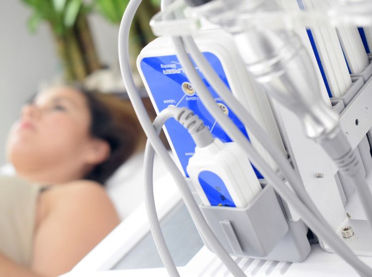
There are many imaging diagnostic methods of breast diseases such as mammography, breast ultrasound, radiotherapy, radioisotope.
- What is the imaging diagnosis
These are techniques of imaging diagnosis such as X-ray (mammogram), breast ultrasound, Magnetic resonance imaging (MRI), SPECT imaging, PET-CT imaging …
Currently, three techniques like breast ultrasound, X-ray mammogram and MRI are popularly used in Vietnam and other countries to diagnose breast diseases and play important roles, especially in screening – early detection of breast cancer.
- What is serious breast disease?
That is breast cancer! It is the top disease in kinds of cancer happened to women. Mortality rate for breast cancer accounts for the highest number of many kinds of cancer in women.
According to the statistics showed in France, approximately 40,000 women are diagnosed with breast cancer and over 10,000 cases died from this disease. It can be cured if it is diagnosed soon.
- How is the value and choice of imaging diagnosis equipment in screening – detection of breast cancer?
Unlike European and American women, there is more fat in the mammary gland than the glandular tissue (fatty breast), most Asian women own thickened mammary gland (more glandular tissue than fatty one). A recent report in Singapore which carried out a survey of women in high risk has showed that the special effect of ultrasound method was 70.7% and X-ray of breast accounted for 75.6%. It proves that mammogram plays an important role in the early detection of breast cancer in Asian women.
Nowadays, common recommendation is that, firstly, using mammography and then, combining with ultrasound will increase the rate of early detection of breast cancer.
Breast MRI is very sensitive (96-100%) and specific (>90%), but so expensive. It is often used when the result of two above methods is uncertain.
- Principles of breast cancer screening guidelines.
The American Cancer Society (ACS) recommends for all women as follows:
➢ Women ages 20 to 39:
- Breast exam by your doctor every three years
- A monthly breast self-exam
➢ Women ages from 40:
- Annual breast cancer screening with mammograms
- Annual breast exam by your doctor
- A monthly breast self-exam
With those who own thickened mammary gland, they should combine X-ray and/or MRI of breast.
Risk factors for breast cancer:
➢ Have a first-degree relative (mother, sister) who are diagnosed with breast cancer:
- If your relative has breast cancer after age 40 or cancer in both breasts, you should to start a routine mammogram from the age of 30
- If a relative has breast cancer before age 40, you should start a routine X-ray of breast at age 10 years before the age of your relatives.
(For examples: your relative has breast cancer at age 36, you should have a mammogram at age 26)
➢ Have a first-degree relative who are diagnosed with ovarian cancer
➢ Other factors:
- Early Puberty (before age 13)
- Late menopause (after age 55)
- Childless or no breastfeeding
- Having first child late (after age 30)
- Get obesity after menopause
- Are the imaging diagnosis harmful to health?
Ultrasound and magnetic resonance are naturally non-radiating; so, they should be completely harmless on machines used in medicine.
Today, mammograms with a small amount of radiation, less than 1mSv/time, which is far fewer than the radiation dose that people absorb annually from the natural environment. This dose is very low in the range of radiation safety; so, it is harmless to people’s health.
- When is the appropriate time for using imaging diagnosis techniques?
Generally, due to the influence of female hormones in the menstrual period, mammary gland becomes denser and more sensitive (painful) in the days before menstrual period. Thus, in order to avoid the false signal due to the hormone impact as well as to reduce the sensitivity when you take the breast examination by imaging techniques, you are advised to have an exam in the first half of menstrual cycle, preferably within 07 to 10 days after the first menstrual period (mainly for X-ray and MRI of breast).
- Do these diagnostic techniques cause any discomfort for patients during the procedure of examination?
When taking the mammogram, you usually stand or sit and your breasts will be compressed lightly between two plates of X-ray machine. In common, you will feel compressed but not pain. This machine usually has automated technology which not only ensures enough/moderate pressure on breast without pain but also meets the requirement of image quality.
In magnetic resonance imaging, you will comfortably lie face down on the table with a special support device, called a coil, in which your breasts are placed and compressed lightly to keep them stable. Completely painless.
In ultrasound procedure, you are required to lie on your back on the ultrasound table. Your hands raised to the top. The transducer gently move to scan over the breasts and both armpits through a soft layer of gel which helps to transmit the sound waves well. This technique will be comfortable and gel will be cleaned off your breast by a normal absorbent paper. This special gel does not irritate the skin.
Dr. Vu Tan Duc – Dean of Department of Imaging Diagnosis, University Medical Center HCMC
Dr. Ho Hoang Thao Uyen, Department of Imaging Diagnosis, University Medical Center HCMC


